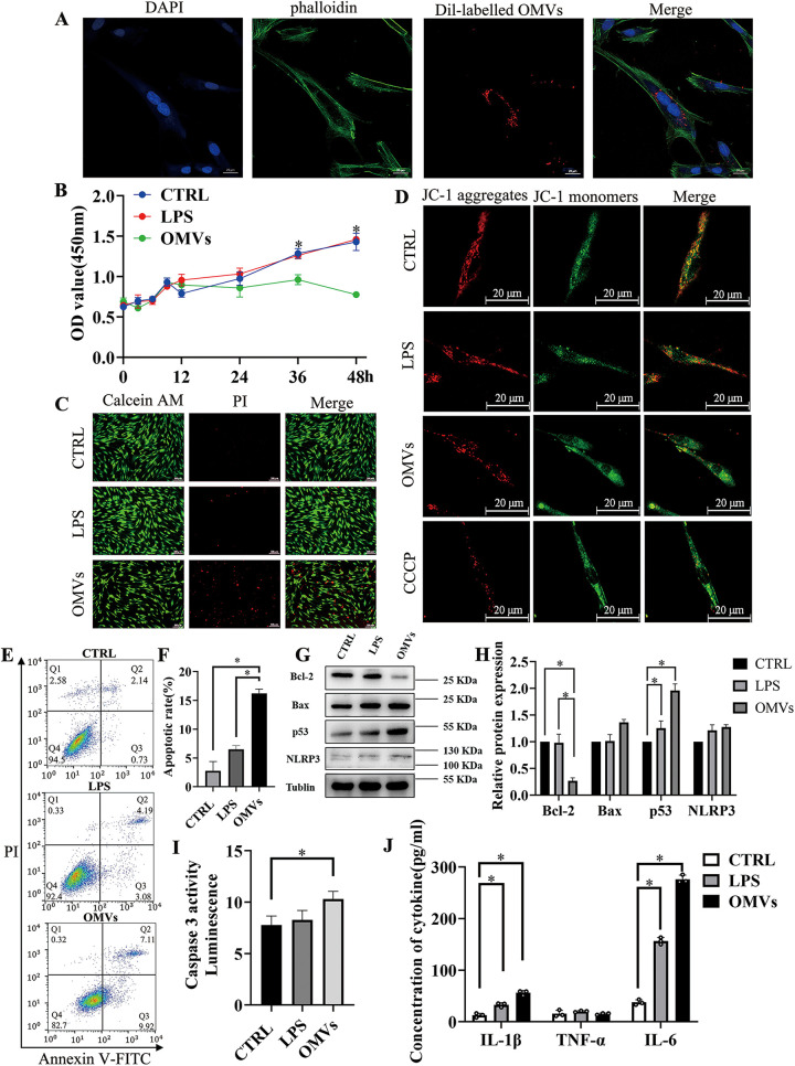
FIG 3.
P. gingivalis OMVs decrease cell viability and promote apoptosis and inflammation in hPDLCs. (A) Endocytosis analysis demonstrated that the isolated P. gingivalis OMVs were taken up by the hPDLCs. (B) Cell proliferation of hPDLCs stimulated by the LPS or OMVs was measured with the CCK-8 assay. (C) PI staining image of control, LPS-treated, or OMV-treated hPDLCs. (D) The fluorescent probe JC-1 was used to measure mitochondrial membrane potential. CCCP, carbonyl cyanide m-chlorophenyl hydrazone. (E and F) Flow cytometry analysis of hPDLCs after being treated with LPS or OMVs. (G and H) Western blotting showed the expression of apoptosis-related proteins like BclII, Bax, p53, and inflammation-associated protein NLRP3 in hPDLCs treated with LPS or OMVs. (I) Caspase 3 activity was measured with the caspase-3 assay kit. (J) Secretion of different cytokines of hPDLCs measured by ELISA. Data are shown as mean ± SD. Data between two groups were compared using Student's t test. Cell experiments were conducted three times independently. *, P < 0.05.

