FIG 1.
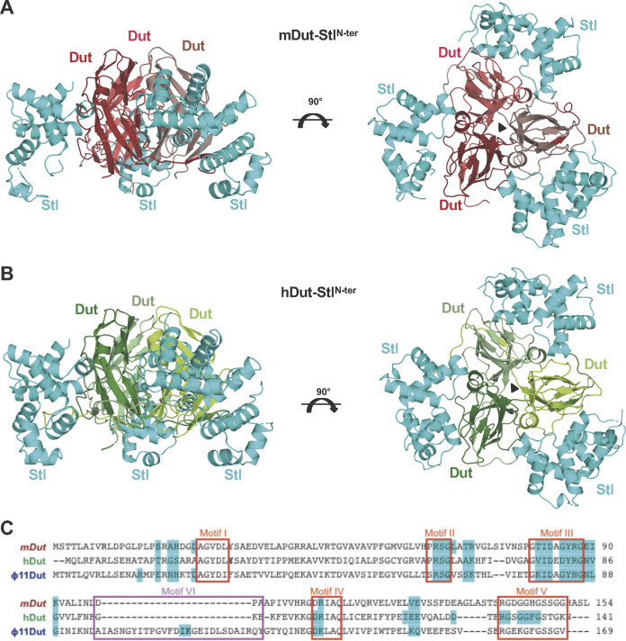
Crystal structures of mDut-StlN-ter and hDut-StlN-ter complexes. (A and B) Cartoon representation of StlN-ter in complex with Duts from M. tuberculosis (A) and human (B). For both complexes, two orthogonal views are shown. Representation scheme: (A) each protomer of trimeric mDut is colored in red and StlN-ter protomers are in cyan; (B) each protomer of trimeric hDut is colored in green and StlN-ter protomers are in cyan. (C) Alignment of mDut, hDut, and ϕ11 Dut. The residues of each Dut which interact with StlN-ter are highlighted in cyan, the catalytic motifs of trimeric Duts are in red boxes, and the S. aureus phage-specific motif VI is in a magenta box.

