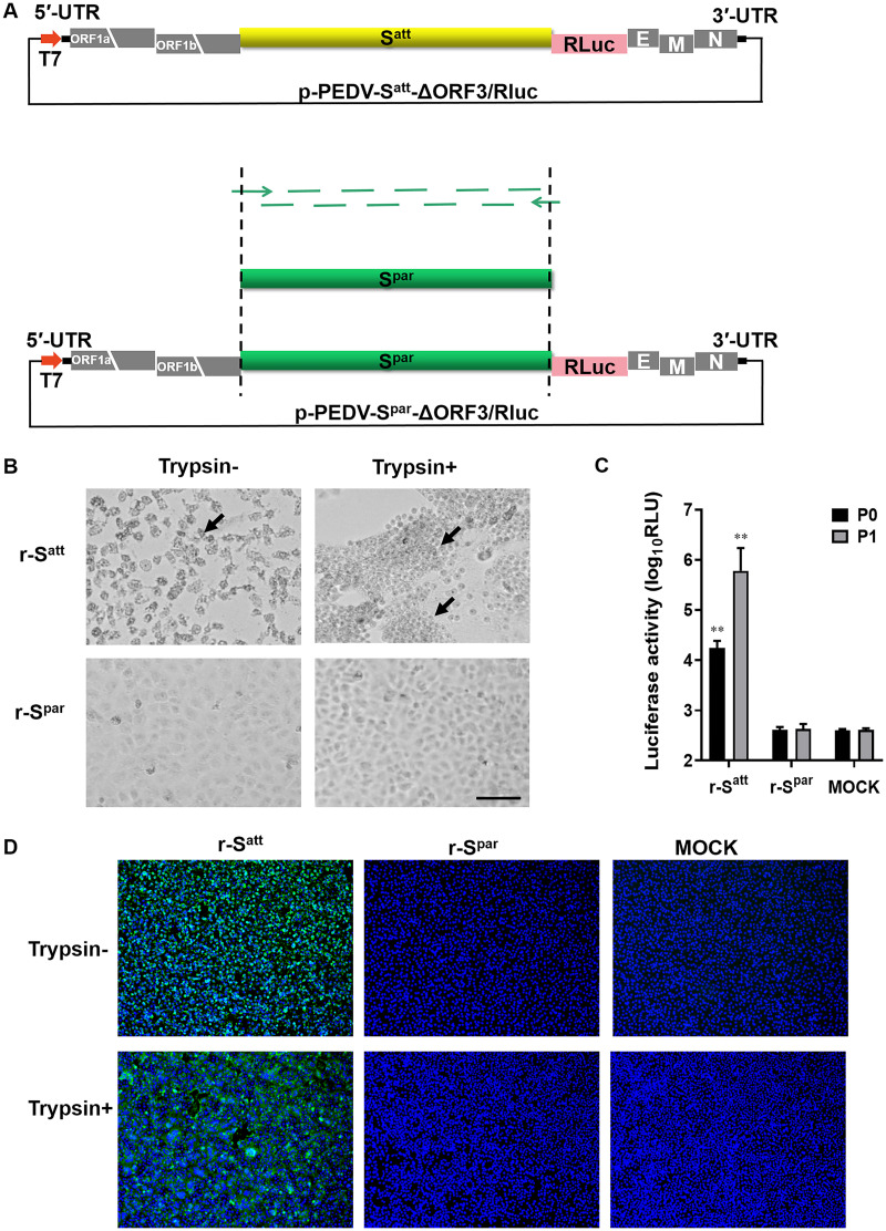
FIG 1.
The S gene determines Vero cell adaptation of PEDV DR13att. The rescue of recombinant viruses (r-Spar and r-Satt) was carried out by using a reverse genetic system based on homologous RNA recombination. Briefly, LR7 cells infected with mPEDV (a recombinant PEDV with the DR13att backbone carrying the ectodomain of the S gene of mouse hepatitis coronavirus, thereby enabling the virus to infect murine LR7 cells) were harvested. Next, capped runoff RNA transcripts (donor RNA) synthesized from the PacI-linearized transfer vectors were transfected into the mPEDV-infected LR7 cells by electroporation. Finally, the electroporated cells were overlaid onto confluent Vero cells and cultured in the presence or absence of trypsin for the rescue of recombinant viruses, and candidate recombinant viruses were purified by two rounds of endpoint dilutions. (A) Construction of the transfer vector p-PEDV-Spar-ΔORF3/RLuc. The transfer vector contains the truncated 1a/1b and the structural genes of the PEDV genome. The thick red arrow indicates the T7 promoter from which donor RNA was synthesized in vitro using T7 RNA polymerase. The S gene of the PEDV DR13 parental strain (Spar) (GenBank accession number DQ862099) was obtained by using fusion PCR with artificially synthesized DNA fragments. p-PEDV-Spar-ΔORF3/RLuc was constructed by replacing the S gene of the PEDV DR13 attenuated strain (Satt) in p-PEDV-Satt-ΔORF3/RLuc with the Spar gene. UTR, untranslated region. (B) CPE of r-Spar and r-Satt on Vero cells. Vero cells in 24-well plates were infected with r-Spar (its culture) and r-Satt in the presence or absence of trypsin (15 μg/mL). The formation of CPE was observed at 36 h postinoculation (hpi). Arrows indicate CPE. Bar, 100 μm. (C) Luciferase expression by r-Spar and r-Satt. The intracellular Renilla luciferase activities of the first two generations (P0 and P1) of recombinant viruses were determined when obvious CPE appeared or at 5 days postinoculation (y axis) (relative light units [RLU]). The results are expressed as the mean values from three parallel tests, and error bars represent the standard deviations (SD). Comparisons were carried out between the luciferase activities of the recombinant viruses and that of the mock treatments. *, P < 0.05; **, P < 0.01. (D) Indirect immunofluorescence assay (IFA) of r-Spar- and r-Satt-infected cells. Vero cells were infected with r-Satt and r-Spar in the presence or absence of trypsin (15 μg/mL). Infected cells were fixed at 36 hpi and immunolabeled with rabbit anti-PEDV M polyclonal antibody (green). Nuclei were labeled with DAPI (blue).

