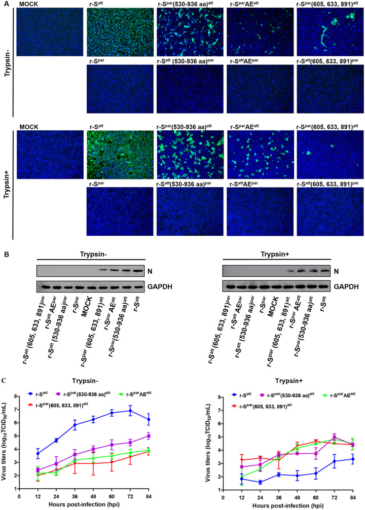
FIG 5.
Characterization of the recombinant PEDVs in Vero cells. (A) IFA of recombinant PEDVs. Eight recombinant PEDVs, r-Spar, r-Satt, r-Satt(530–936 aa)par, r-Spar(530–936 aa)att, r-SattAEpar, r-SparAEatt, r-Satt(605, 633, 891)par, and r-Spar(605, 633, 891)att, were inoculated into Vero cells in the presence or absence of trypsin (15 μg/mL). Cells were fixed at 36 hpi and immunolabeled with rabbit anti-PEDV M polyclonal antibody (green). Nuclei were labeled with DAPI (blue). (B) Western blot analysis of Vero cells infected with recombinant PEDVs. Vero cells inoculated with recombinant PEDVs were harvested at 24 hpi. Proteins in the lysed cells were separated by 12% SDS-PAGE and subsequently processed for immunoblot analysis with monoclonal antibody against the PEDV N protein; GAPDH served as a protein loading control. (C) Multistep growth kinetics of recombinant PEDVs. Vero cells were inoculated with r-Satt, r-Spar(530–936 aa)att, r-SparAEatt, and r-Spar(605, 633, 891)att (MOI of 0.0012) and continued to be cultured in the presence or absence of trypsin (15 μg/mL). At the indicated times postinfection, cells and culture media were harvested by three rounds of freezing and thawing, followed by centrifugation to remove cell debris. The supernatants were collected and used to measure viral titers in the presence of trypsin. Results are expressed as the mean values from three parallel tests, and error bars represent the SD.

