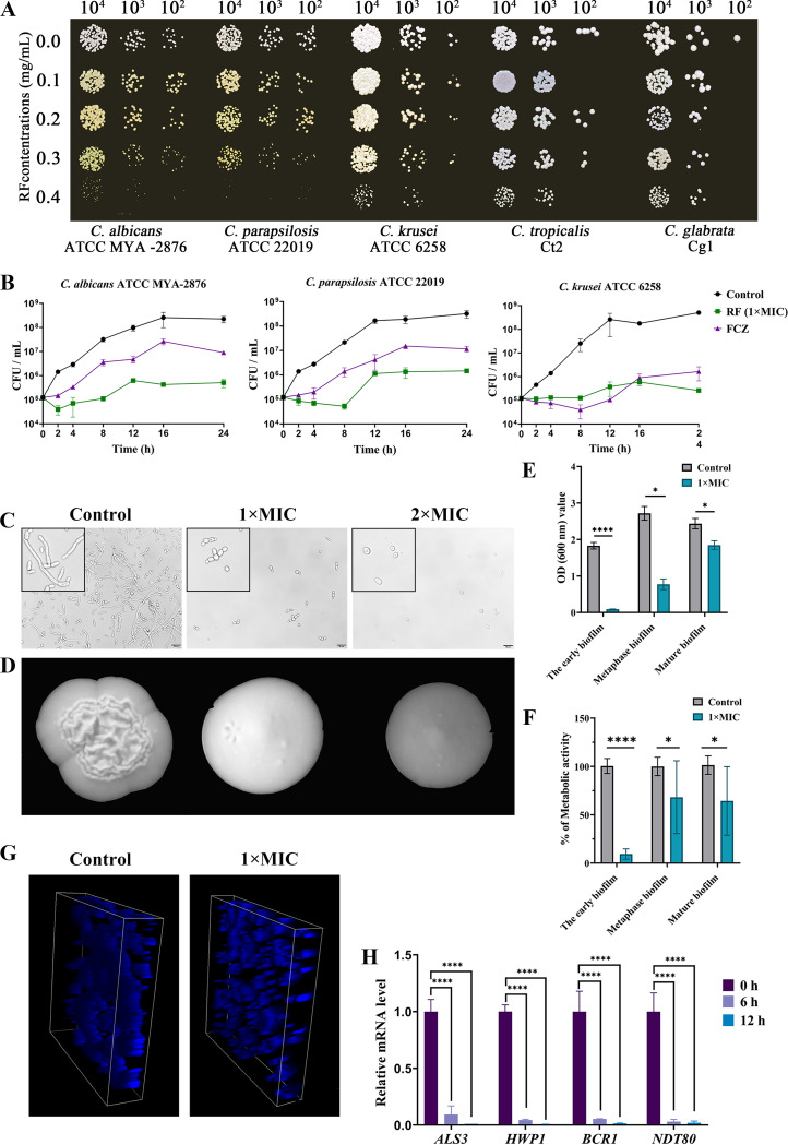
FIG 1.
The inhibitory effect on cell growth and hyphal and biofilm formation of Candida species. (A) The growth of Candida species on YPD agar plates with different concentrations of RF. (B) Growth curves of standard strains C. albicans ATCC MYA-2876, C. krusei ATCC 6258, and C. parapsilosis ATCC 22019 treated with 0.4 mg/mL of RF. (C) Hyphal formation was evaluated in RPMI 1640 plus 10% (vol/vol) FBS liquid medium, and uniformly enlarged images are presented in the black boxes on the left side. Bar, 20 μm. (D) Hyphal formation was evaluated on YPD plus 10% (vol/vol) FBS agar plates. (E) The biomass of C. albicans biofilm was observed by a crystal violet assay. (F) Metabolic activity of C. albicans biofilm was determined by a 2,3-bis-(2-methoxy-4-nitro-5-sulfophenyl)-2H-tetrazolium-5-carboxanilide assay. The results are presented as relative percentages. (G) The three-dimensional structure of C. albicans biofilm was stained with calcofluor white and observed by confocal laser scanning microscopy. (H) The expression of biofilm-related genes was determined. C. albicans treated with 1× MIC of RF was incubated for 0, 6, and 12 h before RNA extraction. 1× MIC, 0.4 mg/mL of RF; 2× MIC, 0.8 mg/mL of RF. Data were analyzed by a t test or one-way analysis of variance (one-way ANOVA) (ns [not significant], P > 0.05; *, P < 0.05; **, P < 0.01; ***, P < 0.001; ****, P < 0.0001).

