FIGURE 6.

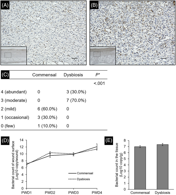
Evaluation of wound inflammation based on the infiltration of neutrophils. Wound tissue was stained with anti‐myeloperoxidase heavy chain antibody to confirm infiltration of neutrophils. The images of granulation tissue in the commensal group (A) and dysbiosis group (B) were shown. Region enclosed in solid box in lower magnification (×4.0, Scale bar = 300 μm) is at higher magnification images (×20, Scale bar = 50 μm). (C) Data show the number of rats assigned to each class. *Wilcoxon rank sum test. (D,E) The bacterial count of the wound surface and wound tissue was determined based on the copy number of tuf gene. Each error bar represents the SE
