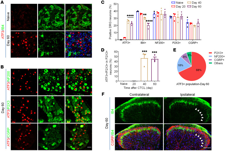
Figure 2. CTCL results in nerve injury in the late phases.
(A) Double immunostaining for ATF3 and IB4 in cervical DRGs of naive and CTCL mice at day 60. Scale bar: 25 μm. (B) Double immunostaining of ATF3 and the neuronal markers NF200, P2X3, and CGRP in cervical DRGs from CTCL mice at day 60. Scale bar: 25 μm. (C) Percentage of ATF3+, IB4+, NF200+, P2X3+, and CGRP+ neurons in the cervical DRG of CTCL mice at different time points. n = 3. Two-way ANOVA, F(12, 30) = 9.15, P < 0.0001. (D) Percentage of ATF3 colocalization with P2X3+ neurons in the cervical DRG of CTCL mice at different time points. n = 3. One-way ANOVA, F(3, 8) = 31.89, P < 0.0001. (E) Pie chart showing ATF3 colocalization with NF200+, P2X3+, and CGRP+ neurons in the cervical DRG from a CTCL mouse on day 60. Data were collected from 3 animals. (F) Double immunostaining for CGRP and IB4 in the cervical spinal cord dorsal horn from CTCL mice at day 60. Arrowheads indicate the loss of IB4+ primary afferents in the IIi. Scale bar: 100 μm. Data are expressed as the mean ± SEM. One- or 2-way ANOVA with Bonferroni’s post hoc test, **P < 0.01, ***P < 0.001, and ****P < 0.0001 versus the naive group.

