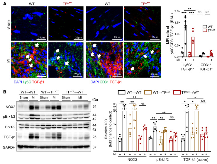
Figure 9. Myeloid cell TF cytoplasmic domain phosphorylation mediates ERK1/2–TGF-β1–dependent cardiac remodeling in permanent LAD ligation.
(A) Confocal microscopy of myocardial cryosections obtained from WT (C57BL/6J) and TFΔCT mice. Representative images and quantification of MFI of Ly6C+TGFβ-1+ and CD31+TGF-β1+ cells. Ordinary 1-way ANOVA, Šidák’s multiple-comparison test; n = 5 animals per group. Scale bars: 25 μm. (B) Mice with transplanted BM were subjected to permanent LAD ligation versus sham surgery and investigated 7 days later. Western blot analysis of NOX2 (normalized to GAPDH), p-ERK1/2 (normalized to total ERK1/2), and TGF-β1 (normalized to GAPDH) in infarcted myocardium obtained from chimeric mice. Ordinary 1-way ANOVA, Šidák’s multiple-comparison test; n = 5–7 animals per group. Data are shown as mean ± SEM. *P < 0.05, **P < 0.01, ***P < 0.001.

