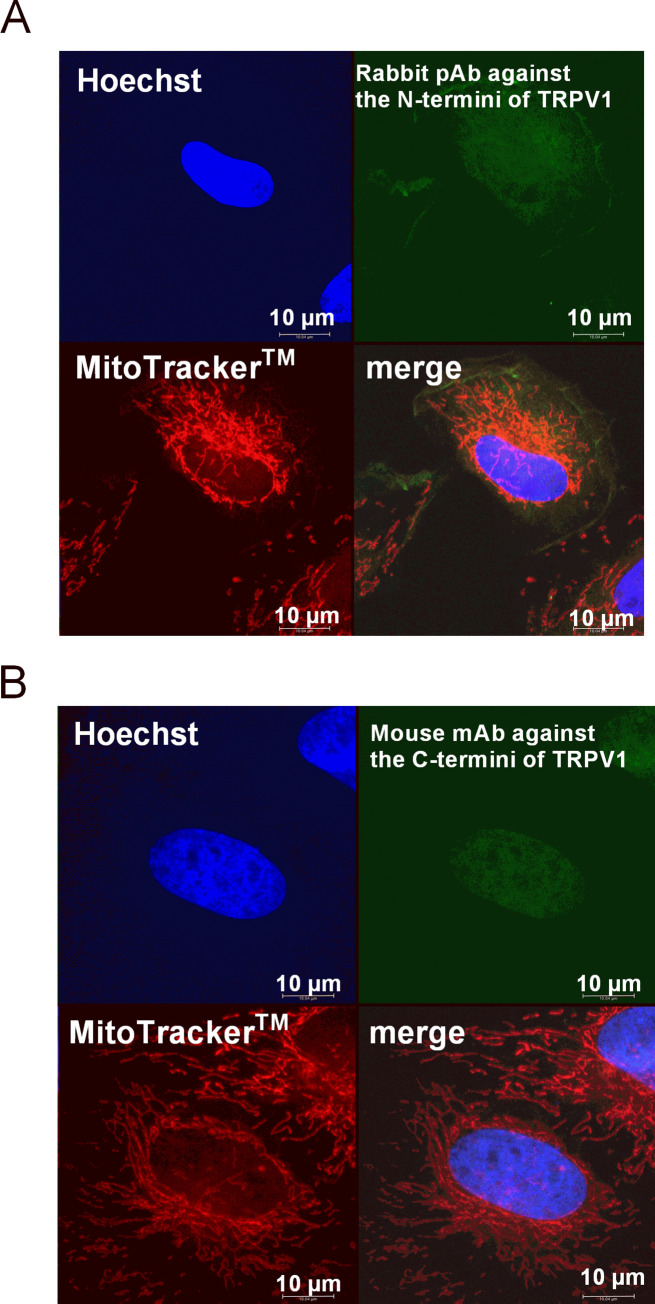
Fig 4. TRPV1 staining in mitochondria-labeled VSMC.
Confocal microscopy images of triple-stained rat VSMCs (objective magnification 63x): Hoechst (blue) for nucleus, FITC-conjugated secondary antibody (green) bound to the (A) polyclonal antibody against the TRPV1 N-terminus or (B) monoclonal antibody against the TRPV1 C-terminus, and MitoTracker® DeepRed FM (red) for mitochondria.

