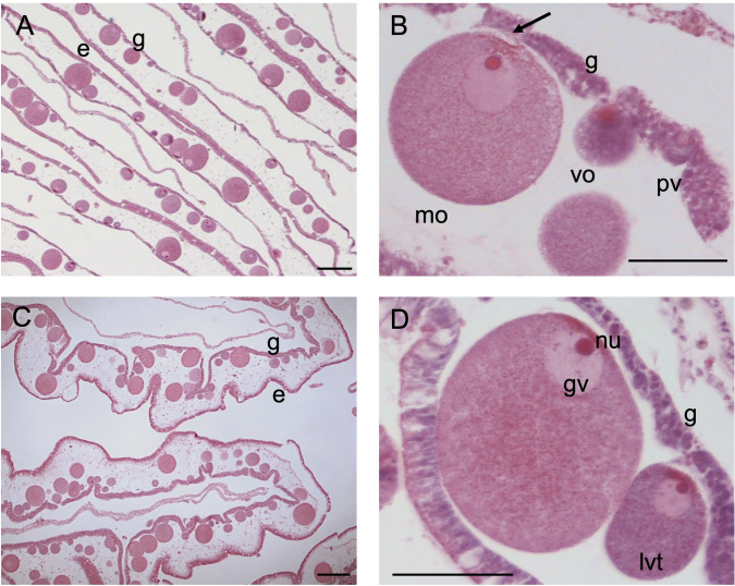
Fig 1. Histological section of a female gonad.
(A, C) Ovary (B, D) Development of oocytes. g gastrodermis, e endodermis, pv pre-vitellogenic oocyte embedded within gastrodermis, vo vitellogenic oocyte, lvt late-vitellogenic oocyte mo mature oocyte, arrow residual linkage with trophocytes (paraovular body), nu nucleolus, gv germinal vesicle. Scale bars: A, C = 100 μm; B, D = 50 μm.

