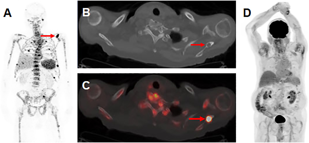
Figure 5:
Visualization of skeletal myeloma by 89Zr-DFO-daratumumab immunoPET in an 80 year-old male with osseous myeloma. (A) MIP image from a 89Zr-DFO-daratumumab PET/CT demonstrates multiple foci of osseous avidity, including a left scapular focus (arrow). (B) Axial CT (C) and fused PET/CT images from the 89Zr-DFO-daratumumab PET/CT demonstrate the left scapular focus localizes to a lytic osseous lesion on CT (arrows). (D) MIP image from an 18F-FDG PET/CT 1 week prior fails to identify the lesions seen on 89Zr-DFO-daratumumab PET/CT.
Image reprinted with permission from the Radiological Society of North America from Ulaner GA, Sobol NB, O’Donoghue JA, et al. CD38-targeted immunoPET of multiple myeloma: from xenograft models to first-in-human imaging. Radiology. doi: 10.1148/radiol.2020192621. In press.

