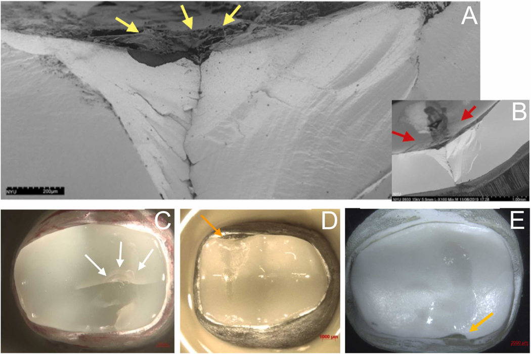
Fig. 6 –
SEM images of a failed glass-infiltrated 5Y specimen (A, B) and stereomicroscope images of representative failed restorations (C,D,E). Higher magnification (A) shows defects on the 5Y surface glass layer (yellow arrows). In B, the relationship between failure and occlusal wear scar (red arrows). which resulted in a cone crack failure. C shows cracks and a quasiplastic deformation at the load application area (white arrows) in a lithium disilicate restoration. Edge chipping fracture (orange arrows) in a glazed 5Y (D) and in a silica-infiltrated 3Y (E).

