
Figure 1.
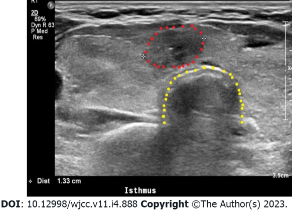
Thyroid ultrasonography. Diffusely enlarged thyroid gland with rounded lobes showing the diffusely heterogeneous and coarse echotexture of the thyroid gland. The isthmus nodule was suspected to be malignant owing to its ill-defined margin. The red dots indicate the isthmus nodule; this nodule was identified as a papillary carcinoma by fine-needle aspiration cytology. The yellow dots indicate the trachea.