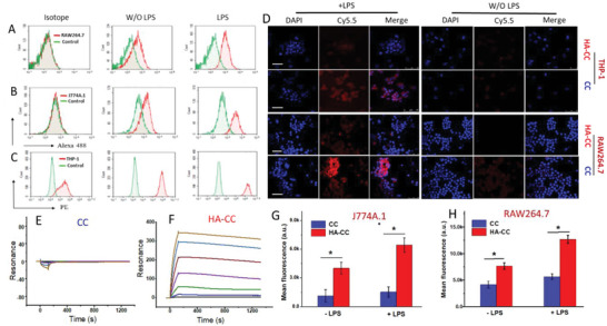
Figure 2.

A–C) Flow cytometric analysis of CD44 expression in macrophages following LPS stimulation. D) Confocal laser scanning microscopy analysis of enhanced cellular internalization of HA‐CC in THP‐1 and RAW264.7 cells with or without 24 h LPS stimulation. Non‐targeted CC was the control. Scale bar: 50 µm. Improved hyaluronic acid‐modified cerasome (HA‐CC) macrophage interaction with or without 24 h LPS stimulation. SPR analysis of the binding between CD44 and CC F) with or E) without HA conjugation. Flow cytometric analysis of G) J774A.1 and H) RAW 264.7 cells after incubation for 2 h with fluorescent Cy5.5‐labeled HA‐CC (HA‐CC‐Cy5.5) or fluorescent Cy5.5‐labeled CC (CC‐Cy5.5). Differences among the groups were determined using ANOVA with Tukey's multiple comparison test (n = 5), and *p < 0.05 was considered significant.
