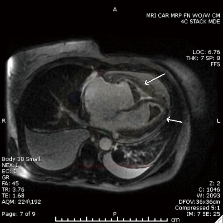
Figure 2.

Cardiac MRI with delayed post-contrast 2D showing a thrombus within the left ventricular apex, right ventricular apex, and right ventricular outflow tract, with generalized subendocardial enhancement throughout the right and left ventricles (arrows). MRI, magnetic resonance imaging; 2D, two-dimensional.
