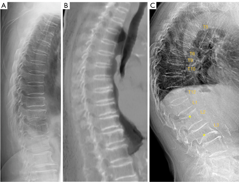
Figure 4.
Differentiation of multiple aSVs and multiple osteoporotic vertebral fractures. (A) A female case radiograph from the current study, showing multiple aSVs. (B) A female case from another study (sagittally reconstructed CT scan), showing multiple aSVs. (C) A female case radiograph from another study, showing multiple OVFs. In (C), OVFs vary greatly in shape and severity, and most show endplate depression. * on L2 shows upper endplate fracture (depression); * on L3 show lower expansive endplate (anomaly). In (A,B), multiple aSVs show much less variation in shape and severity, and aSVs do not show apparent wedging or endplate depression. In (B), the increased density of the involved endplates suggests regenerative inflammatory changes. aSVs, acquired short vertebrae; CT, computed tomography; OVF, multiple osteoporotic vertebral fracture.

