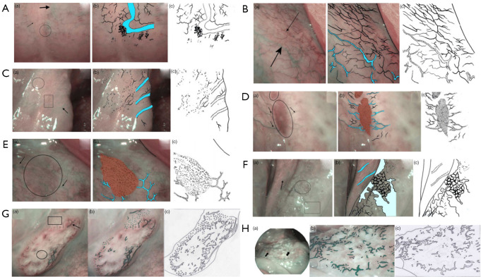
Figure 2.
The classification raised by Tirelli. (A) Normal pattern in type 1 epithelium: (a) posterior hard palate appearance on NBI endoscopy. Superficial mucosal capillaries (thin arrow), submucosal veins (thick arrow), and flower-like structures (circle); (b) superficial mucosal capillaries, submucosal veins (in cyan), and flower-like structures; (c) schematic drawing. (B) Normal pattern in type 2a epithelium: (a) floor of mouth appearance on NBI endoscopy. Superficial mucosal capillaries (thin arrow), submucosal veins (thick arrow); (b) superficial mucosal capillaries and submucosal veins (in cyan); (c) schematic drawing. (C) Normal pattern in type 2b epithelium: (a) inferior trigone appearance on NBI endoscopy. Superficial mucosal capillaries (circle, rectangle), submucosal veins (arrow); (b) superficial mucosal capillaries and submucosal veins (in cyan); (c) schematic drawing. (D) Dysplastic pattern in type 1 epithelium: (a) severe dysplasia of the hard palate on NBI endoscopy. A well-demarcated brownish/purple area with thick dark spots is visible (circle); it is perpendicularly reached by dilated light blue vessels (arrows); (b) the well-demarcated brownish/purple area perpendicularly reached by dilated light blue vessels (in cyan); (c) schematic drawing. (E) Dysplastic pattern in type 2a epithelium: (a) severe dysplasia of the floor of mouth on NBI endoscopy. A well-demarcated brownish/purple area with thick dark spots is visible (circle); it is perpendicularly reached by dilated light blue vessels (arrows); (b) the well-demarcated brownish/purple area perpendicularly reached by dilated light blue vessels (in cyan); (c) schematic drawing. (F) Dysplastic pattern in type 2b epithelium: (a) severe dysplasia of an inferior trigone on NBI endoscopy. Mesh (circle), leukoplakia (rectangle) and the submucosal vessels (arrow); (b) the image resembles a mesh, leukoplakia, and the submucosal vessels (in cyan); (c) schematic drawing. (G) Neoplastic pattern in an ulcerated cancerous lesion: (a) ulcerated cancer of the hard palate on NBI endoscopy. Necrosis in the center of the lesion (circle), dark green spots (rectangle), dilated winding vessels or bobby-pin (arrow); (b) dark green spots, dilated winding vessels, or bobby-pin; (c) schematic drawing. (H) Neoplastic pattern in a vegetating cancerous lesion: (a) vegetating cancer of the floor of mouth on NBI. Unstructured vessels appearing as dark green spots, dilated winding vessels or as bobby-pin (arrow); (b) dark green spots, dilated winding vessels, or bobby-pin; (c) schematic drawing. This figure is licensed by John Wiley and Sons. NBI, narrow-band imaging.

