Figure 3.
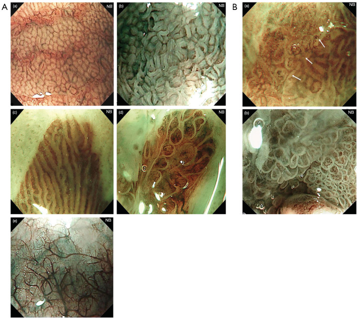
High resolution magnification endoscopy plus NBI findings of Barrett’s epithelium. (A) Narrow-band endoscopic imaging of in non-dysplastic Barrett’s esophagus shows microstructural and microvascular changes: (a) RMSP with round pattern; (b) RMSP with tubular pattern; (c) RMSP with linear pattern, (d) RMSP with villous pattern; (e) absent microstructural pattern. (B) Narrow-band endoscopic imaging of in neoplastic Barrett’s esophagus shows microstructural and microvascular changes: (a) figure shows a clear demarcation line (white arrows) between the neoplastic mucosa with irregular microvessels (left part) and the non-neoplastic mucosa with regular microstructural pattern (right part); (b) figure shows IMSP with irregular microvascular pattern. This figure is licensed by John Wiley and Sons. NBI, narrow-band imaging; IMSP, irregular microstructural pattern; RMSP, regular microstructural pattern.

