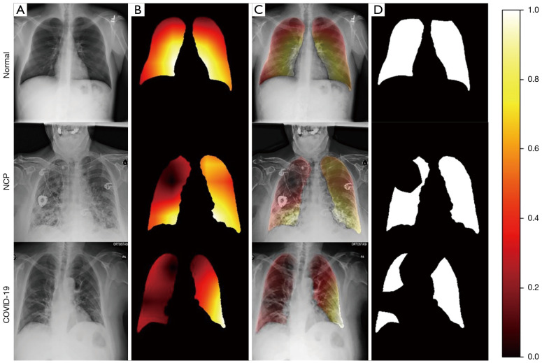
Figure 4.
Representative cases of (A) original chest Radiograph images; (B) lung region activation maps generated by the proposed network; (C) lung region activation maps overlaid on the chest Radiograph images; (D) binary mask of the region of interest. Colors represent the importance scores, which represent the importance of each pixel in a feature map towards the overall classification decision. The lung region which has a value greater than 0.2 is segmented as the ROI. From top to bottom, the three cases were from normal class, non-COVID-19 pneumonia class and COVID-19 class respectively. NCP, non-COVID-19 pneumonia; COVID-19, coronavirus disease 2019; ROI, region of interest.

