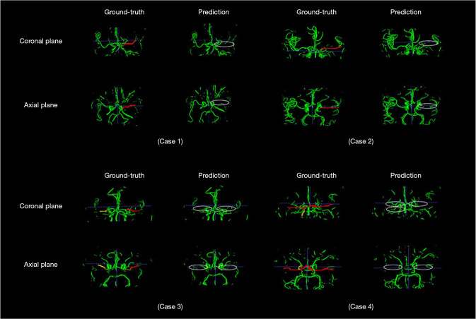
Figure 4.
Vessel segmentations of two patients with unilateral stenosis and two with bilateral stenosis. The red and yellow in ground truth, represent severe and mild stenosis/occlusion labeled or supplemented by experts, and the green is the normal vessels. The ellipses in prediction contain the stenosis or occlusion lesions predicted and voted by the proposed model and post-processing method.

