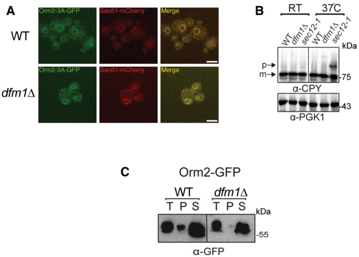
Figure EV4. Orm2‐3A accumulates exclusively in the ER. Related to Fig 7 .

- Fluorescence imaging was performed as in Fig 7B except for WT and dfm1∆ cells expressing Orm2‐3A‐GFP was used. Sec61‐RFP (ER marker, red) was used to test for co‐localization with Orm2‐3A‐GFP (2 biological replicates; n = 2). Arrowheads indicate Orm2 co‐localizing in post‐ER compartments. Scale bar, 5 μm.
- dfm1∆ cells do not abrogate COPII‐mediated export of CPY. The indicated cells were either grown at room temperature or shifted to non‐permissive growth at 37°C. Cells were analyzed by SDS–PAGE and immunoblotted for CPY with α‐CPY and PGK1 with α‐PGK1.
- Western blot of aggregated versus soluble Orm2‐GFP at the ER. Lysates from WT and dfm1Δ cells containing ORM2‐GFP were blotted using anti‐GFP to detect Orm2. P is ER aggregated fraction and S is ER soluble fraction.
