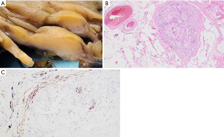
Figure 3.
Lifting agent granuloma in a 65-year-old male (Case 3). (A) Gross pathology specimen, cross section of colonic wall with lesion. (B) Sub-serosal blood vessel infiltration by lifting agent granuloma (hematoxylin & eosin, 40×). (C) Positive smooth muscle actin immunohistochemical staining highlighting the muscular wall of sub-serosal blood vessel involved by lifting agent granuloma (immunohistochemistry, 100×).

