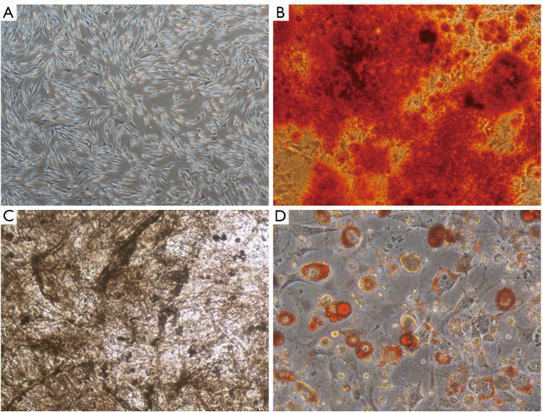
Figure 2.
Characterization of BMSCs. (A) Morphology of BMSCs observed by light microscope (×40). (B-D) Differentiation capability of BMSCs into osteoblasts and adipocytes evaluated by Alizarin Red S staining (B: ×100), alkaline phosphatase staining (C: ×100) and Oil Red O staining (D: ×200). BMSCs, bone marrow mesenchymal stem cells.

