Figure 2.
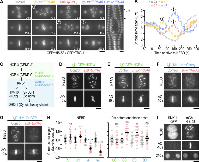
PP4 inhibition diminishes outer but not inner kinetochore assembly prior to NEBD. (A) Selected images and kymograph from time-lapse movies of one-cell embryos co-expressing GFP::histone H2B (HIS-58) and GFP::γ-tubulin (TBG-1). Time is relative to NEBD. Scale bar, 5 µm. (B) Chromosome span versus time relative to NEBD (mean of n embryos ± SEM). Measurements were performed in time-lapse movies such as those shown in A. Numbers in A and B refer to corresponding time points in images and graph. (C) Assembly hierarchy of the kinetochore components analyzed in this figure and Fig. S2. (D–G) Selected images from time-lapse movies of one-cell embryos co-expressing fluorescently tagged kinetochore components (GFP::HCP-3, GFP::HCP-4, and HIM-10::GFP are endogenously tagged; KNL-1::mCherry is a functional transgene) and transgenic GFP- or mCherry-tagged histone H2B (HIS-58). Only the kinetochore component is shown. Time points correspond to NEBD and the last frame prior to anaphase onset (AO). Scale bars, 5 µm. (H) Intensity of the chromosomal signal at NEBD or just prior to AO in the one-cell embryo (mean ± 95% CI), normalized to the mean of the respective control, for the components shown in D–G. Statistical significance was determined by the Mann-Whitney test. ****P < 0.0001; **P < 0.01; ns = not significant, P > 0.05. (I) Selected images from a time-lapse movie of a one-cell embryo co-expressing endogenously tagged SMK-1::GFP and transgenic mCherry::histone H2B (HIS-58). Scale bar, 5 µm.

