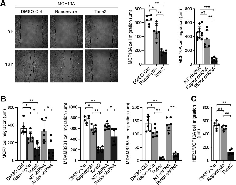
FIGURE 1:
mTORC2 promotes the migration of MCF10A and breast cancer cells. Wound closure cell migration assays were performed in the presence of rapamycin, Torin2, or 0.1% DMSO control (Ctrl); and with cells previously treated with shRNAs to silence raptor or rictor, or a nontargeting (NT) shRNA as control, as described in the Materials and Methods section. (A) MCF10A cell migration. Representative images of MCF10A cells in the wound closure assay are shown on the left, taken immediately after wounding and addition of the indicated treatments (0 h) and after 18 h. (B) Cell migration data for MCF7, MDAMB231, and MDAMB453 cells. (C) HER2/MCF10A cell migration. Graphs show the measured migration distances ±SD of at least six wounds from at least three independent experiments. *, p < 0.05; **, p < 0.01; ***, p < 0.001.

