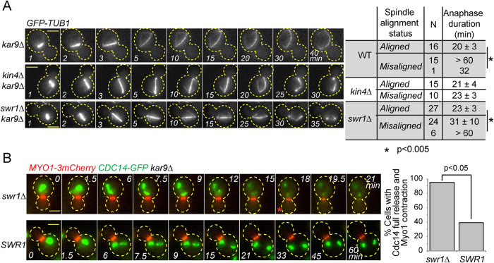
FIGURE 5:
Deletion of SWR1 causes slippage from the SPOC arrest. (A) Duration of anaphase in indicated cell types during correct spindle alignment and spindle misalignment. GFP-labeled tubulin (GFP-TUB1) was monitored by time-lapse microscopy with 1-min time resolution. The duration of anaphase was calculated as the time elapsed from the start of the fast spindle elongation phase (metaphase-anaphase transition) until spindle breakdown. N, number of cells analyzed. Asterisks indicate a significant difference according to Student’s t test (p < 0.05). Representative still images of indicated GFP-TUB1 kar9∆ cells monitored during spindle misalignment are shown on the left. Cell boundaries are outlined with dashed lines. Arrows indicate the point of spindle breakdown. Time point 1 is 1 min before the metaphase-anaphase transition, which coincides with the start of fast spindle elongation phase. Scale bar: 3 µm. (B) Representative still images from the time-lapse movies of CDC14-GFP MYO1-3mCherry bearing kar9∆ cells with misaligned spindles. Scale bar: 3 µm. The red asterisk indicates the onset of Cdc14 full release. The percentage of cells with misaligned spindles that released Cdc14-GFP fully and contracted Myo1-3mCherry is plotted. Time-lapse series of 21 cells were analyzed each in SWR1 and swr1∆. A chi-square test was performed using a significance level of 0.05. Please note that a higher percentage of kar9∆ Myo1-3mCherry cells with misaligned spindles released Cdc14 and exited mitosis in comparison to kar9∆ cells shown in A. This is most likely a consequence of Myo1 tagging, as it was not observed in cells with untagged Myo1.

