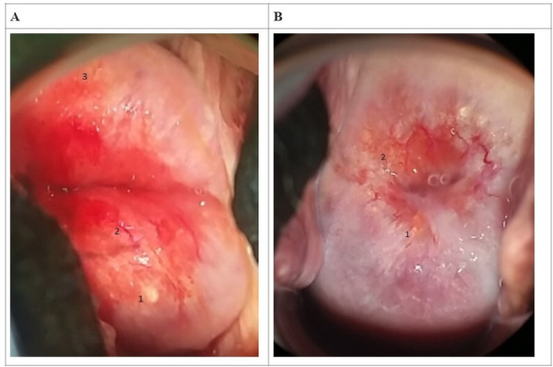
Figure 2.

Imagery of female genital schistosomiasis pathology of the cervix with reported lesions (magnification of ×4). (A) 1—rubbery papule, 2—abnormal blood vessel, 3—homogeneous sandy patch (30-year-old woman, +UGS, +lower abdominal pain, +menstrual irregularity). (B) 1,2—grainy sandy patches (45-year-old woman, −UGS, +lower abdominal pain, +menstrual irregularity).
