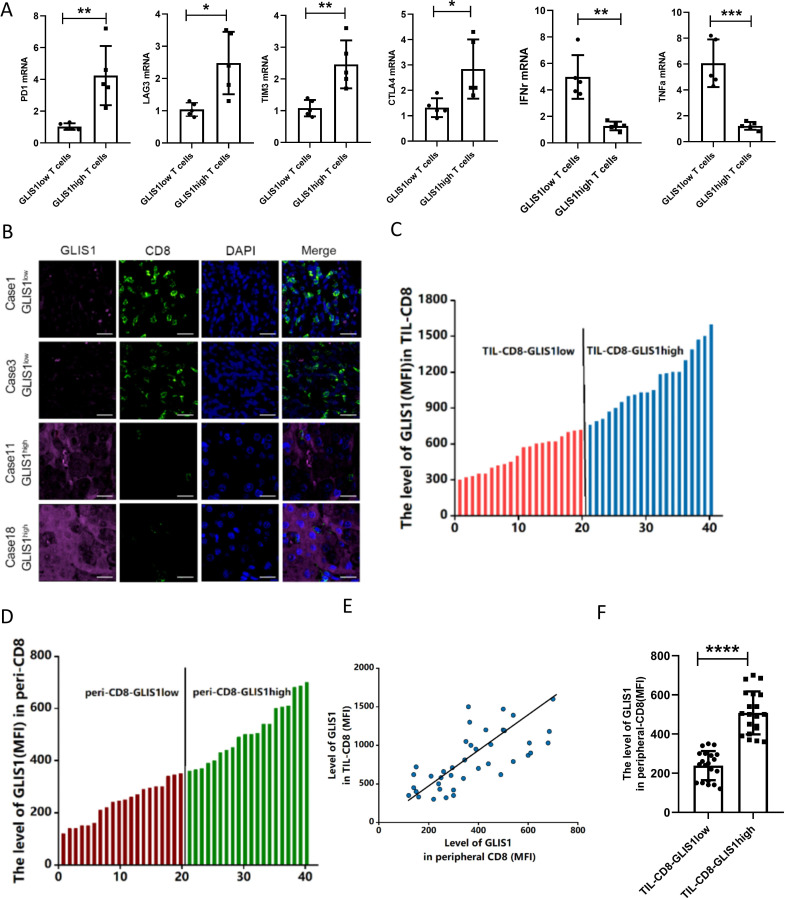
Figure 2.
The expression of GLIS1 in PBMC CD8+ T cells of HCC patients predicted the immune status in tissues. (A) GLIS1 expression in CD8+ TILs from 10 HCC patients tissues was analyzed by flow cytometry, and were divided into high GLIS1 expression and low GLIS1 expression groups. The qRT-PCR results of the common markers of CD8+ T cell exhaustion (PD1, LAG3, TIM3 and CTLA4) and cytokines (IFN-γ, TNF-α) expression in each group. (B) Samples from HCC patients CD8+ TILs were used for immunofluorescence detection of CD8 and GLIS1 expression in each group. (C, D) The expression of GLIS1 in CD8+ TILs and corresponding peripheral blood CD8+ T cells of 40 HCC patients were analyzed by flow cytometry, respectively, and ranked them from low to high. (E, F) The correlation analysis between the GLIS1 expression in CD8+ TILs and PBMC CD8+ T cells. *p<0.05, **p<0.01, ***p<0.001, ****p<0.0001. HCC, hepatocellular carcinoma; PBMC, peripheral blood mononuclear cell; qRT-PCR, quantificational real-time PCR; TILs, tumor infiltrating lymphocytes.

