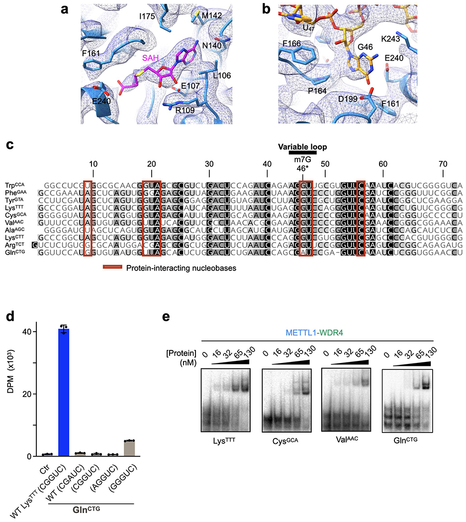
Extended Data Fig. 6: Structural and sequence organization of tRNA.

a-b, Sharpened cryo-EM map (mesh) near the SAH-binding site (a) and the G46 binding pocket (b). c, Sequence alignment of the human tRNAs used in this study shaded by conservation. Red boxes indicate nucleobases within 4 Å of protein. Sequences were aligned using Clustal Omega and visualized by Geneious Prime. d, In vitro methylation activity of full-length METTL1-WDR4 for the indicated tRNAs with the specified variable loop sequences, shown as mean ± SD from 3 replicates. e, EMSA using METTL1-WDR4 with different tRNAs shows no dramatic differences in affinities. Representative images from 3 replicate experiments are shown. For each gel, protein concentrations are 0, 16, 32, 65, and 130 nM, left to right.
