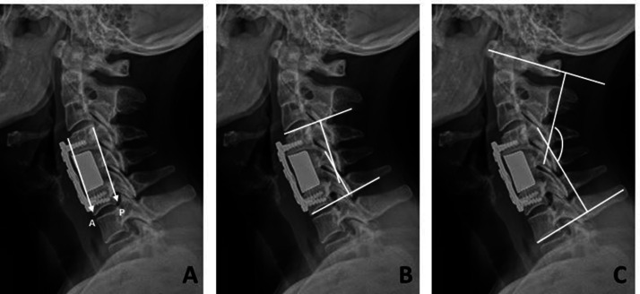
Figure 1.
(A) Implant subsidence was assessed on lateral radiographs at the cranial and the caudal end plates of the upper and the lower vertebrae in the affected segment. The distance between the upper end plate of the upper vertebral body and the lower end plate of the lower vertebral body was measured at the anterior and the posterior points of the end plate. Severe subsidence was defined as a loss of height of >3 mm. (B) Segmental sagittal alignment was defined as the angle between the cranial and the caudal end plates of the upper and the lower vertebrae in the affected segment. (C) Cervical lordosis was measured using Cobb angle from C1 to C7. A, anterior segment height; P, posterior segment height.

