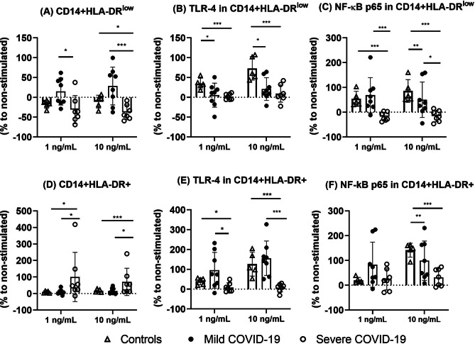
Fig. 4.
The frequencies of CD14 + HLA-DR.low (A) monocytes expressing TLR-4 (B) and NF-κB (C) and CD14 + HLA-DR + (D) expressing TLR-4 (E) and NF-κB (F) in ex vivo LPS-stimulated whole blood of healthy controls and mild and severe COVID-19 patients. Whole blood samples of controls, mild COVID-19, and severe COVID-19 patients were aliquoted in tubes to a volume of 1 mL and incubated (5%CO2, 37 °C) with two different LPS concentrations (1 and 10 ng/mL, Sigma-Aldrich, USA) or without immunogenic stimulus for 2 h. The phenotype of monocytes was evaluated by flow cytometry. The frequencies of each cell subtype were evaluated by the % of difference compared to the non-stimulated condition. *(p < 0.05), **(p < 0.01), and ***(p < 0.001) denote statistical difference (p < 0.01)

