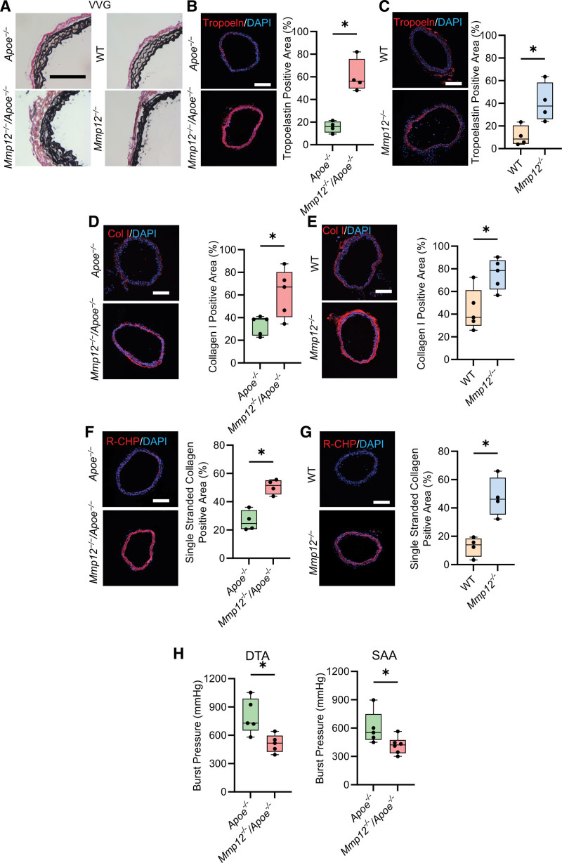
Figure 3.
Effect of Mmp12 deficiency on aortic wall extracellular matrix composition. A, Representative examples of Verhoeff–van Gieson (VVG) staining of SAA in Apoe-/- and Mmp12-/-/Apoe-/- (left column) and WT and Mmp12-/- (right column) mice. Scale bar: 250 µm. B through G, Representative images of immunofluorescence staining and quantification of tropoelastin (red, B and C), collagen type I (red, D and E) and single-stranded collagen using R-CHP (red, F and G) in Apoe-/- and Mmp12-/-/Apoe-/- (B, D, and F) and WT and Mmp12-/- (C, E, and G) mice. Nuclei are stained blue with DAPI. n=4 per group. Scale bar: 500 µm. H, Burst pressure, as a functional readout of wall strength, of DTA and SAA in Apoe-/- and Mmp12-/-/Apoe-/- mice. n=5 to 6 per group. *P<0.05, **P<0.01 by 2-tailed Mann-Whitney U test (B through H). DAPI indicates 4′,6-diamidino-2-phenylindole; DTA, descending thoracic aorta; MMP, matrix metalloprotease; R-CHP, collagen hybridizing peptide, Cy3 conjugate; SAA, suprarenal abdominal aorta; and WT, wild-type.

