Fig. 1.
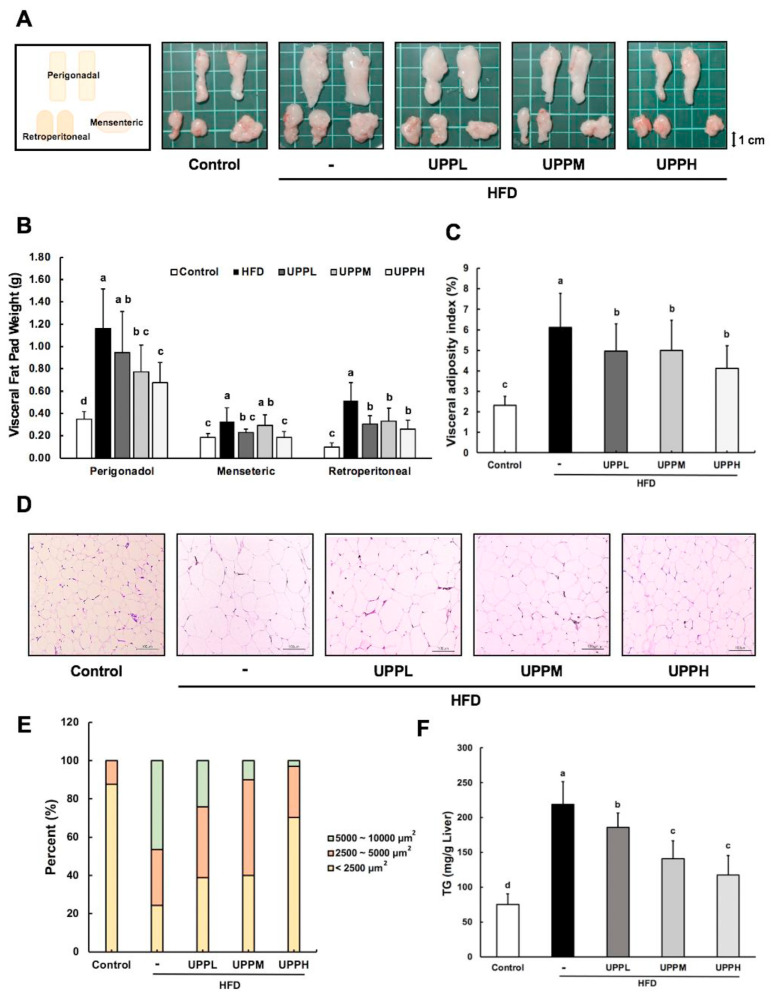
Effect of Ulva prolifera polysaccharide on epididymal white adipose tissue and liver in high-fat diet-fed mice.
A) Representative images of visceral fat pads (epididymal white adipose tissue) from each group of mice are shown at the end of week 15. Perigonadal, retroperitoneal, and mesenteric fat pad, respectively, are depicted. B) Visceral fat pad weights were measured after 15 weeks of treatment. C) Visceral adiposity index (%) was calculated as {[perigonadal + mesenteric + retroperitoneal fat pad weight (g)]/BW (g) * 100}. D) Representative images of hematoxylin and eosin (H&E)-stained perigonadal fat pads of mice in each group. Scale bar, 100 μm. Data are expressed as mean ± SD (B–C, n = 11 mice/group). E) Adipocytes size in visceral adipose tissue was determined by Image J. F) Hepatic triglyceride levels. Scale bar, 100 μm. Data are expressed as mean ± SD (C, n = 8 mice/group). The statistical significance of differences among the five groups were analyzed by one-way ANOVA and Duncan’s multiple range tests. The values with different letters (a–d) are significantly different ( p < 0.05) between each group.

