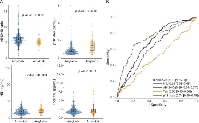
Figure 1. Dot-Plots Distributions of Blood Biomarkers According to Amyloid-PET Status and ROC Curve Analysis.
(A) Boxplots indicate median, 1st and 3rd quartiles; the black diamonds indicate the mean. p-values are for nonparametric Wilcoxon rank tests. (B) ROC curve analyses showing the performance of the 4 blood biomarkers to discriminate amyloid positivity on PET. Aβ = amyloid beta; AUC = area under the curve; NfL = neurofilaments light chain; ROC = receiver operating characteristic.

