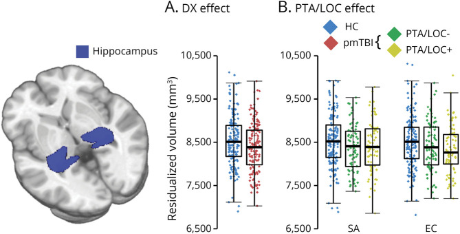
Figure 3. Group Effects in Subcortical Volume Analysis.
Panel A presents subcortical volumetric results for primary analyses examining differences between diagnoses (DX: healthy controls [HC] = blue diamonds; patients with pediatric mild traumatic brain injury [pmTBI] = red diamonds). Panel B presents pmTBI subgroups based on injury characteristics (no reported posttraumatic amnesia or loss of consciousness [PTA/LOC-] = green diamonds; reported PTA/LOC = yellow diamonds). A glass brain rendering of the hippocampus (blue) is present to the left of the panels. All data points in box-and-scatter plots have been residualized (Resid.) to remove the effects of total intracranial volume and age. Hippocampal volumes were reduced in pmTBI relative to HC at both visits (Panel A), but recovery was greater for pmTBI who reported no PTA/LOC at the time of injury. eFigure 4 (links.lww.com/WNL/C525) depicts data for secondary subcortical volumes.

