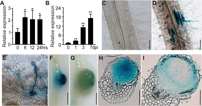
Fig 2. RPG expression pattern in L. japonicus roots.
(A-B) qRT-PCR analysis of RPG transcript levels in roots of wild type (WT) L. japonicus. Samples were collected at 0, 6, 12, and 24 h after inoculation with purified Nod factor (A) or at 0, 1, 3 and 7 days after inoculation with M. loti R7A (B). Expression is relative to that of mock-treated samples (0 h or 0 dpi) and normalized to L. japonicus Ubiquitin. Asterisks indicate significant differences between the mock and the rhizobial/Nod factor treatments at the indicated time points by Students t-test. (C-G) pRPG:GUS expression patterns in L. japonicus roots and nodules. The constructs were expressed in wild type and the transgenic roots were stained with X-Gluc (blue). No GUS was detected in the absence of rhizobia inoculation (C). Strong GUS staining was detected in epidermal cells (D) and (E) and young nodules (F), but there was much lower staining in mature nodules (G). Bacteria were stained by magenta (purple) to indicate ITs (E). (H-I) Nodule sections showed that pRPG:GUS expressed in all cell layers of young nodules (H), but was only expressed in epidermal and nodule parenchyma cells in mature nodules (I). Scale bars: 100 μm (C-E and H-I); 1 mm (F-G).

