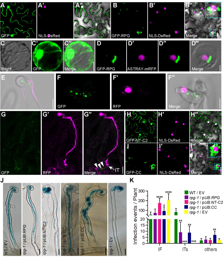
Fig 4. Subcellular localization of the RPG protein in N. benthamiana leaves and L. japonicus roots.
(A, B, H and I) Confocal microscopy images of N. benthamiana leaves expressing p35S:GFP (A), p35S:GFP-RPG (B), and the separate RPG domains p35S:GFP-NT-C2 (H) and p35S:GFP-CC (I). In each, the green, magenta and merged images are shown in adjacent panels. The nucleus was labeled with NLS-DsRed (magenta). Sections within an image that are outlined in dotted Cyan lines are showed enlarged in the top right corner of that image. (C -D) p35S:GFP (C) or p35S:GFP-RPG (green) and the nuclear marker, ASTRAY5-mRFP (magenta) (D), were co-expressed in L. japonicus root protoplasts using a DNA-PEG-calcium transfection method. (E to G) pRPG:GFP-RPG was introduced into rpg-1 by A. tumefaciens-mediated stable transformation. RPG subcellular localization were analyzed using whole-mount immunolocalization with anti-GFP primary antibody and Alexa Fluor 488-conjugated Affinipure donkey anti-Mouse IgG secondary antibody, 5 or 10 days after inoculation with M. loti MAFF303099/RFP. Green shows RPG subcellular localization and magenta shows M. loti. Close arrowheads indicate RPG subcellular localization and arrow indicate IT (G). (J-K) Assays of complementation of the rpg-1 mutant by the predicted NT-C2 domain (pUB:NT-C2) and by RPG lacking the NT-C2 domain (pUB:CC) showing the NT-C2 domain is not required for complementation of infection. Infection phenotypes (J) and Infection events (K) of the pUB:NT-C2 or pUB:CC were expressed in rpg-1, and the phenotypes were scored after inoculation with M. loti R7A/LacZ (n>14). Asterisks indicate significant differences between the transformants with the pUB-RPG constructs and those with the empty vector (EV) (Students t-test). Scale bars: 25 μm (A-B and H-I); 10 μm (C-G); 20 μm (J).

