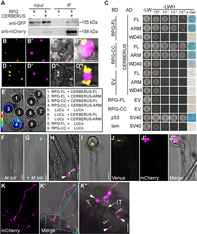
Fig 5. RPG interacts with CERBERUS.
(A) Co-Immunoprecipitation (Co-IP) assay showing the interaction between GFP-RPG and CERBERUS-mCherry in N. benthamiana leaves. GFP-RPG and CERBERUS-mCherry were co-expressed in N. benthamiana leaves. Co-IP was assayed using anti-mCherry antibody, and the precipitated proteins were detected by immunoblot analysis with anti-mCherry and anti-GFP antibodies. One representative result out of two biological replicates is shown. (B and D) BiFC assays of full-length RPG and CERBERUS (B) or RPG-CC and CERBERUS-ARM (D) (yellow) and NLS-DsRed (magenta) in N. benthamiana leaves. The image shows strong Venus fluorescence localized in puncta, some of which were close to the nucleus. (B‴ and D‴) shows an enlargement of the area in outlined in cyan in the merged image (B″ and D″). (C) A GAL4-based yeast two-hybrid system was used to analyze the interaction between CERBERUS and full-length RPG (RPG-FL) or RPG lacking the NT-C2 domain (CC), and between CERBERUS ARM or WD40 and RPG-FL or RPG-CC. Potential interactions were assayed by growth on SD/-LWH (medium without histidine, leucine, or tryptophan) after gradient dilution. Images show the growth of co-transformants on selection media after three days. (E) Luciferase biomolecular complementation assays of the interaction between RPG and CERBERUS or RPG-CC and CERBERUS-ARM in N. benthamiana leaves. The indicated constructs were co-expressed in N. benthamiana leaves, and luciferase complementation imaging was conducted two days after agroinfiltration. LUCn, N-terminal fragment of firefly luciferase. LUCc, C-terminal fragment of firefly luciferase. Fluorescence signal intensity is indicated. (F-G) Live cell confocal images of RPG-CERBERUS BiFC construct was expressed in L. japonicus hairy roots. Venus fluorescence (yellow) was detected in L. japonicus root hairs before rhizobia inoculation (F), and in curled root hairs after rhizobia inoculation (G). (H-K) a RPG-CERBERUS BiFC construct was expressed in M. truncatula sunn-1 by hairy root transformation. The transgenic roots were observed seven or ten days after inoculation with Sm1021/mCherry. mCherry (magenta) shows the nucleus (H) or Sm1021/mCherry (J, K). Close arrowheads indicate nucleus; Open arrowheads indicate Venus fluorescence produced by RPG interacting with CERBERUS; IT: Infection threads. Scale bars: 25 μm (C-D); 10 μm (F-J); 50 μm (K).

