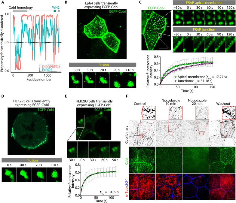
Fig. 2. Cobl underwent LLPS in an MT-dependent manner in cells.
(A) Prediction of internally disordered regions within Cobl. The results of DISOPRED3 (http://bioinf.cs.ucl.ac.uk/psipred) and PrDOS (https://prdos.hgc.jp), which are popular applications that accurately predict internally disordered regions, are shown. Higher propensity scores indicated an increased likelihood that the residue is intrinsically disordered. Considering that a cutoff of 0.5 is frequently used to determine intrinsically disordered residues, Cobl had an abundance of regions that were likely to be intrinsically disordered. (B) In Eph4 cells, EGFP-Cobl condensates underwent fusion within seconds (see also movie S3). Scale bar, 10 μm (low magnification) and 0.5 μm (high magnification). (C) The fluorescence intensities of EGFP-Cobl condensates recovered rapidly, with t1/2 values of 17.27 s (apical membrane) and 31.18 s (junction) (see also movie S4). Scale bar, 10 μm (low magnification) and 0.5 μm (high magnification). N = 4, each. (D) In HEK293 cells, exogenous EGFP-Cobl formed membrane-bound condensates (see also movie S5). Scale bar, 10 μm (low magnification) and 0.5 μm (high magnification). (E) The fluorescence intensities of EGFP-Cobl condensates recovered rapidly, with a t1/2 of 10.09 s. Scale bar, 10 μm (low magnification) and 0.5 μm (high magnification). N = 4. (F) Treatment with 2 μM nocodazole dissolved Cobl condensates in WT cells. Scale bar, 10 μm. Points represent means, and error bars represent SDs.

