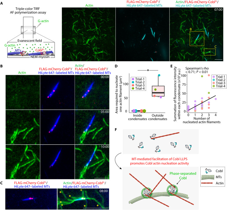
Fig. 8. MT-mediated Cobl LLPS resulted in MT-dependent promotion of Cobl actin nucleation.
(A) Representative TIRF micrographs at 7 min, obtained using triple-color TIRF microscopic analyses (see also movie S12). Protein concentration: Alexa Fluor 488–labeled G-actin, 1 μM; FLAG-mCherry-CoblFL, 100 nM; and HiLyte 647–labeled MTs, 4 μM. Scale bar, 10 μm. (B) MTs elongated AFs directly via CoblFL condensates (see also movie S13). Scale bar, 5 μm. (C) Large Cobl condensates could elongate several AFs, as indicated by yellow asterisks (see also movie S14). Scale bar, 5 μm. (D) The area required to nucleate one AF. *P < 0.05 (Mann-Whitney U test). N = 4 trials, each. (E) Correlation between the number of AFs from each condensate and the fluorescence intensity within each condensate. (F) Schematic drawing of the mechanism underlying MT-mediated facilitation of Cobl actin nucleation activity. In a box plot, solid lines represent medians, boxes represent interquartile ranges, and error bars extending from the box represent the range of data within 1.5 times the interquartile range.

