
Figure 3.
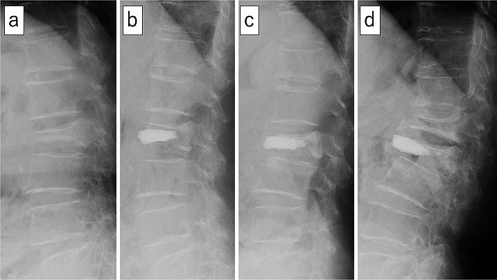
A 78-year-old woman with AVF after augmented vertebral body height loss following BKP.
(a) Preoperative radiograph (lateral view) showing the L2 vertebral fracture (vertebral body height=10 mm).
(b) Postoperative radiograph (lateral view), 1 week after BKP, showing cancellous bone remaining around the cement in the vertebral body.
(c) Postoperative radiograph (lateral view), 1 month after BKP, showing loss of augmented vertebral body height (RAV).
(d) Postoperative radiograph (lateral view), 6 months after BKP, showing an adjacent vertebral fracture at L1.