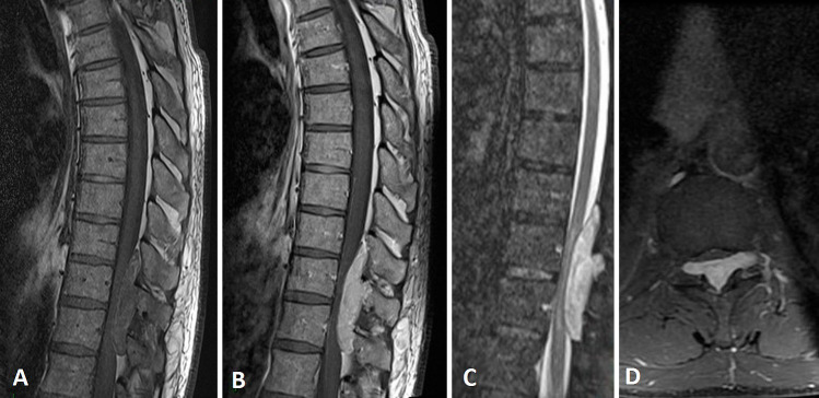
Figure 1.
Preoperative sagittal magnetic resonance imaging of the spine shows an atypical extradural posterior lesion at the level of T10-T11 compressing the cord, which is isointense on T1-weighted image (A), homogenously enhanced with gadolinium (B), and hyperintense on short tau inversion recovery (C). The tumor extended into lateral recesses bilaterally at the level of T10-T11 as shown on the axial view (D).

