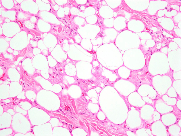
Figure 3.

Photomicrograph of the excision specimen showing a hibernoma with the typical features of a prominent mixture of adipocytes of fetal type. Numerous multivacuolated adipocytes and smaller polygonal cells with abundant cytoplasm are noted, which are characteristic of typical hibernoma (hematoxylin and eosin stain, original magnification 200×).
