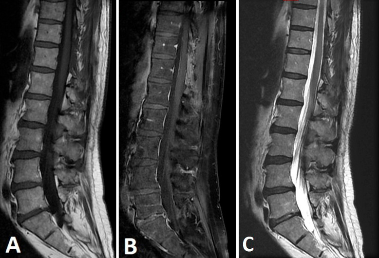
Figure 4.

Postoperative sagittal T1-weighted image before (A) and after gadolinium (B), and T2-weighted (C) magnetic resonance imaging of the spinal cord at the 6-month follow-up showing the absence of any residual contrast uptake or medullary compression.
