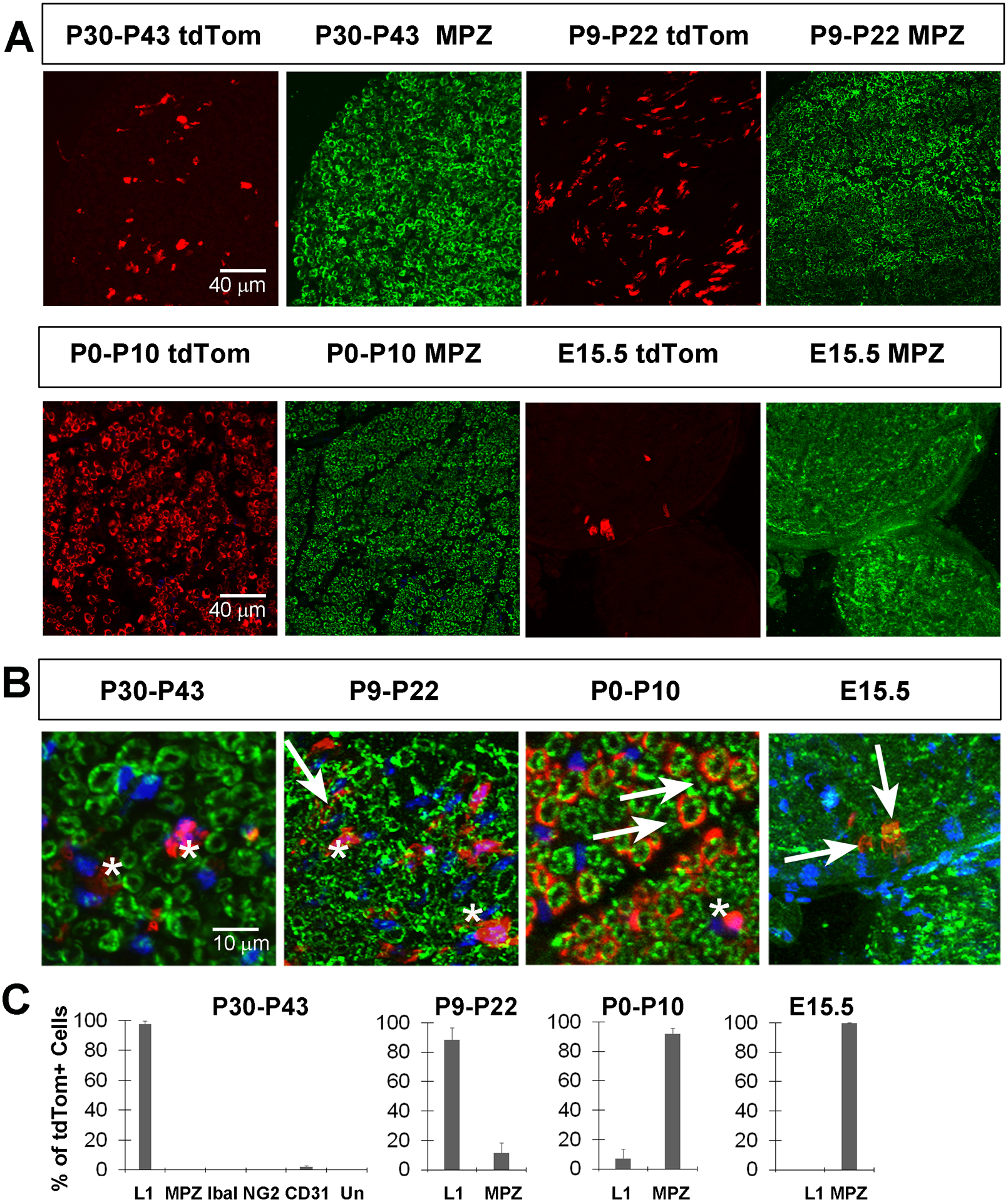
Figure 5. Kir4.1-tdTomato mice given TMX around the onset of myelination or before the end of myelination onset exhibit tdTom+ cells in MPZ+ myelinating Schwann cells (MSC).

A, Images of sciatic nerve cross sections from P50 Kir4.1-tdTomato mice given TMX from P30-P43 (left two panels, top row) or from P9-P22 (right two panels, top row) show tdTom+ cells (red) that fail to co-localize with MPZ+ MSC (green). In contrast, most tdTom+ cells co-localize with MPZ when TMX is given from P0-P10 or from E14-E15. B, High-magnification merged images of tdTomato+ cells and MPZ+ myelinating SC after each of the four TMX treatment regimes. Arrows show double-positive cells and asterisks show tdTom+, MPZ− cells. C, Percentage of tdTom+ cells in sciatic nerve at P50 that express L1, MPZ, Iba1, NG2 or CD31 in mice given TMX from P30-P43 or that express L1 or MPZ in mice given TMX from P9–22, P0-P10 or at E15.5; data in text.
