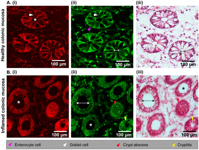
Figure 2.
Comparison of multimodal and H&E images of healthy (A) and inflamed (B) colonic mucosa. The CARS (Ai, Bi) and TPEF (Aii and Bii) images in comparison with H&E stained (Aiii and Biii) images. The asterisks (*) indicate intact and enlarged cryptal lumen regions for healthy and inflamed colonic mucosa, respectively. Arrowhead labels are indicated below the images, and the double-sided arrows measure the lumen diameter for healthy (~ 22 μm) and inflamed colonic mucosa (~ 103 μm).

