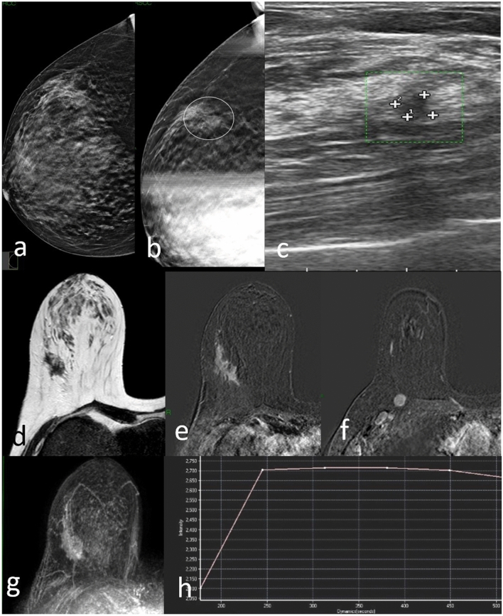
Fig. 2.
DBT right craniocaudal (a) and DBT spot compression (b) tomosynthesis mammogram shows an area of architectural distortion with pleomorphic microcalcifications involving the upper outer quadrant (circle). US image shows suspected hypoechoic area (c). MRI T2W TSE image detects an irregularly shaped mass with spiculations in upper right breast corresponding to the mammographic image (d). Post-contrast subtracted axial fat-suppressed T1-weighted image (e) shows heterogeneous internal enhancement pattern and there is also an ipsilateral prepectoral lymphadenopathy (f). The right breast finding at maximum intensity projection (MIP) image (g); Dynamic MR image reveals a type II T/SI kinetic curve with a rapid initial rise followed by a plateau in the delayed phase (h). Biopsy resulted invasive ductal carcinoma at histological examination

