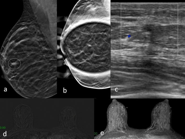
Fig. 3.
DBT right mediolateral oblique projection a shows an area of architectural distortion with a millimeter lump, in the upper periareolar area of right breast (circle). Digital tomosynthesis spot compression view b shows only the architectural distortion, the nodular formation is no longer visible. Focused US image of the right upper periareolar area shows irregular hypoechogenic mass corresponding to mammographic finding (c). No abnormal enhancement observed in dynamic contrast-enhanced MRI study in subtraction T1-weighted (d) and MIP (e). Biopsy resulted ductal carcinoma in situ at histological examination

