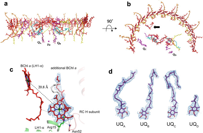
Fig. 2. Arrangement of the cofactors in the Rba. capsulatus LH1–RC.
a Side view along the membrane plane with the periplasm above and the cytoplasm below. Color scheme: BChl a, red sticks; spheroidenes (group-A), yellow sticks; spheroidenes (group-B), orange sticks; BPhe a, magenta sticks; UQ-10, purple sticks; non-heme Fe, magenta sphere. b Top view from periplasmic side of the membrane with the same color scheme as in (a). One additional BChl a and two additional ubiquinones (QC and QD) were detected in the LH1–RC structure. Arow indicates the extra BChl a. c Close contacts (<4.0 Å) between the extra BChl a and surrounding residues, and Mg–Mg distance between the extra BChl a and the nearest LH1-BChl a. Color scheme as in Fig. 1a except for myristate in gray. d All four of ubiquinones (QA, QB, QC and QD) were detected in the LH1–RC structure. The density maps are shown at a contour level of 5.0σ.

