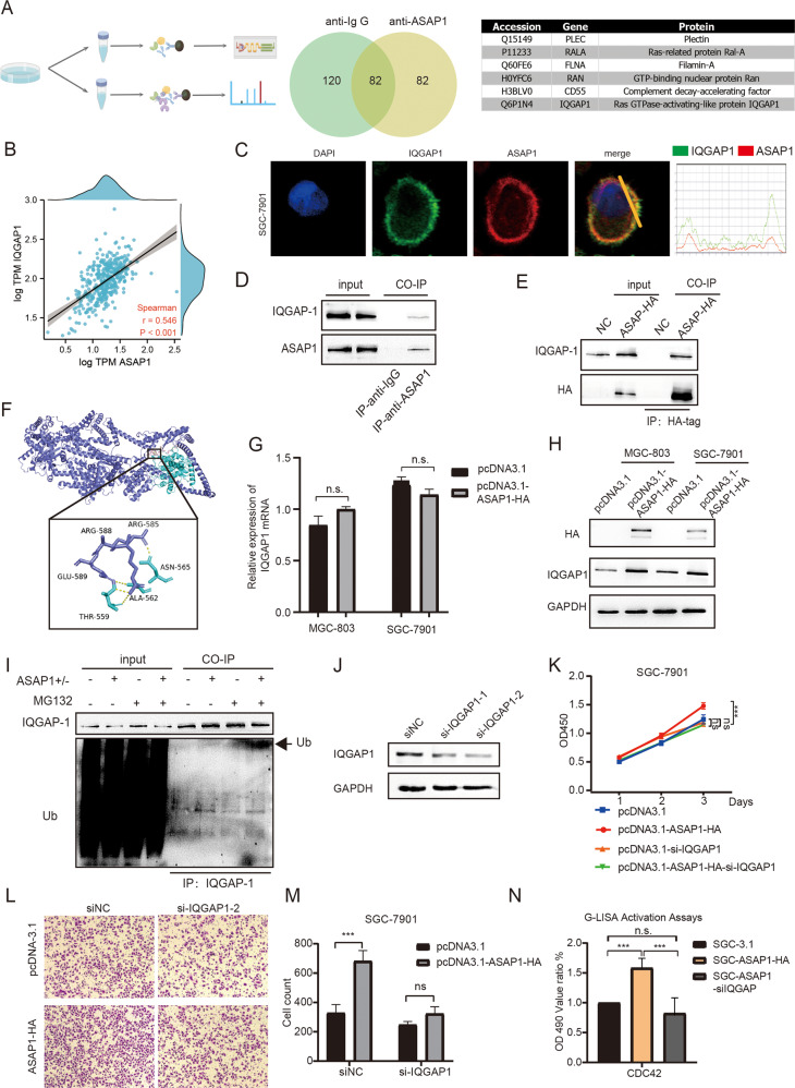
Fig. 6. ASAP1 enhanced CDC42 by interacting with and inhibiting ubiquitin-mediated degradation of IQGAP1.
A The ASAP-associated interactomes were determined by immunoprecipitation and mass spectrometry (IP-MS). Venn diagrams show the number of proteins that may interact listed in the tables. B Correlation analysis of ASAP1 and IQGAP1 RNA expression in TCGA STAD database. C Images showing the immunofluorescence of ASAP1 (red) and IQGAP1 (green). Nuclei were counterstained with DAPI (blue). C SGC-7901 was transfected with pcDNA3.1 or pcDNA3.1-ASAP1-HA. 24 h after transfection, HA-tag fused proteins were immunoprecipitated with their interacting proteins. Western blot was used to detect indicated proteins. D Western blot showing that endogenous ASAP1 co-immunoprecitated with IQGAP1 from SGC-7901 cells. An anti-ASAP1 antibody was used for IP. Normal mouse IgG was used as a control. E Western blot showing that HA-tagged ASAP1 coimmunoprecipitated with IQGAP1 from SGC-7901 cells expressing HA-tagged ASAP1, but not from control SGC-7901 cells. An anti-HA antibody was used for IP. F Molecular docking pattern diagram of ASAP1 and IQGAP1. G IQGAP1 mRNA level of MGC-803 and SGC-7901 after ASAP1 overexpression. H IQGAP1 protein level of MGC-803 and SGC-7901 after ASAP1 overexpression. I SGC-7901 and SGC-ASAP1+/− cells were incubated with MG132 (+) or DMSO (−) for 4 h. Equal amounts of protein lysates were loaded directly onto gels or immunoprecipitated (IP) with an anti-IQGAP1 antibody. Proteins were analyzed by SDS–PAGE and immunoblotting (IB) using anti-ubiquitin and anti-IQGAP1 antibodies, respectively. J Western blot showing that IQGAP was depleted by two different siRNA oligos in SGC-7901 cells. K The proliferation of various types of SGC-7901 cells was determined via CCK-8, including cells transfected with pcDNA3.1, cells transfected with pcDNA3.1-ASAP1-HA, cells transfected with pcDNA3,1 followed by transfection with a siRNA oligo against IQGAP1, and cells transfected with pcDNA3.1-ASAP1-HA followed by transfection with a siRNA oligo against IQGAP1. M The migration of various types of SGC-7901 cells described in L was determined was determined by transwell assays. N The who cell lysates were extracted from various types of SGC-7901 cells to measure CDC42 GTPase activity by G-LISA activation assay, including cells transfected with pcDNA3.1, cells transfected with pcDNA3.1-ASAP1-HA, and cells transfected with pcDNA3.1-ASAP1-HA followed by transfection with a siRNA oligo against IQGAP1.

